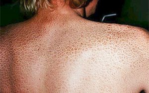
Ichthyosis Vulgaris is a skin disease commonly known as fish skin disease. Constantly dying skin cells accumulate on the skin, causing images like fish scales.
Diseased genes in the mother and father cause this disease to occur in the baby. Symptoms of the disease can sometimes be observed immediately after birth. Sometimes it is noticed in childhood.
Dry skin structure, formations in the form of fish scales, thickening of the skin are listed. While these thickenings are usually on the elbows and knees, they may sometimes spread to the arms and hands.
Keratinization is the transformation of the underlying epidermal cells into hard, shield-like corneocytes that cause the uppermost stratum corneum to act as a barrier.
Scaling is the result of the failure of the corneocytes to separate from the skin, which is the final stage of keratinization. Thus, scaling is seen as a result of the accumulation of corneocytes that cannot be shed. Any keratinization disorder may occur congenitally or acquired.
Scaling can cause significant physical and psychological disturbances. Xeroderma is simply known as dry skin and is literally flaking of the skin. It can be seen as a result of diseases that cause keratinization disorder. Xeroderma is common in atopic eczema with old age and worsens on low humidity winter days.
Xeroderma treatment brings showering instead of bathing with frequent use of moisturizers and moisturizing cleansers.
Autosomal dominant ichthyosis is characterized by thin and diffuse scaling, which is worse on the extensor sides of the body. Keratosis pilaris may accompany and there are keratin plugs in the hair follicle channels in this disease. Its frequency is 0.2%, and its biochemical cause is not known exactly. Moisturizers and, if necessary, keratolytic agents can sometimes be helpful.
The scaling in X-linked ichthyosis is more prominent. Affected male children are often born postmature and difficult. The metabolic disorder is mainly due to steroid sulfatase deficiency, which can be detected in fibroblast, epidermal and lymphocyte cultures. Women are carriers because it is X-linked recessive and its frequency is 1 in 6000.
Non-bullous ichthyosiform erythroderma is rare. It is autosomal recessive and is characterized by diffuse erythema and fine scaling. Although oral acitretin has serious side effects, it may help some patients.
In the rare bullous ichthyosiphroma erythroderma (also known as epidermolytic hyperkeratosis), it tends to diffuse erythema, folded hyperkeratosis, and bullae. There are characteristic reticular degenerative changes in the upper epidermis. The disorder is autosomal dominant and there are known keratin gene mutations. Oral retinoids improve appearance but can also increase vesicles.
Lamellar ichthyosis is a rare autosomal recessive disease with severe hyperkeratosis and scaling.
Patients with non-bullous ichthyosiform erytoderma, epidermolytic hyperkeratosis, and lamellar ichthyosis may be born with a collodion membrane. This bright transparent membrane peels off after a week. Rarely, it can occur in normal babies. Since the barrier function of the skin is impaired, they need careful care.
It can be seen after acquired ichthyosis, lymphoma, human immune deficiency virus (HIV) disease, leprosy, essential fatty acid deficiency and the use of some lipid-lowering drugs.
Darier’s disease is rare and may be autosomal dominant or sporadic. Brownish scaly papules can be seen on the upper body and face. Palmar pits and nail dystrophy also occur. Histologically, suprabasilar separation and premature keratinization are evidence.
Tuberous sclerosis is rare and is an autosomal dominant inherited disease with many anomalies. Pink red papules around the nose and cheeks are characteristic.
Von Recklinghausen disease (neurofibromatosis) is a rare autosomal dominant disease. The development of pinkish soft tumors is observed. Brown macules “cafe au lait” spots may accompany this condition.